
Blogs

Blogs

Myoglobin: Powering Muscles, One Oxygen Molecule at a Time.

Myoglobin is an iron-containing protein found mainly in the muscle cells of vertebrates, particularly in the heart and skeletal muscles. The main function of Myoglobin is to store oxygen and make it available to muscles when energy demands are high. This is especially important during strenuous exercise or prolonged periods of limited oxygen availability, such as deep diving in marine mammals like whales and seals. In terms of structure, myoglobin resembles a single subunit of haemoglobin. However, myoglobin is a monomer, whereas haemoglobin is made up of four subunits. Myoglobin also binds oxygen more strongly than haemoglobin, allowing it to take up oxygen from haemoglobin and supply it to muscle tissues when needed.

Clinical Significance of Myoglobin and Interpretation

Its clinical importance is associated with its use in identifying and monitoring various medical conditions, such as:

  • Early Detection of Myocardial Infarction: Myoglobin rises faster and appears in serum within 2-4 hours of a heart attack.
  • It is used to identify skeletal muscle destruction, which is termed Rhabdomyolysis.
  • It is used to monitor muscle injury from accidents, surgery or muscular diseases.
  • Myoglobin is also detectable in the urine as a result of hereditary myopathies.

Different Diagnostic ways to test for myoglobin levels

A doctor recommends a myoglobin test to measure myoglobin levels in serum or plasma.

The normal range for myoglobin levels is <115 ng/mL in healthy people. Commonly used methods to detect myoglobin levels in laboratories include fluorescence, chemiluminescence immunoassay (CLIA), peroxidase method, creatine kinase test, colloidal gold method, and immunoturbidimetric method.

The chemiluminescence immunoassay are used to quantitatively measure myoglobin in human serum or plasma as an aid in the diagnosis of myocardial infarction.

Conclusion Myoglobin provides oxygen to muscles, allowing them to maintain high activity for extended periods. When muscle damage occurs, myoglobin from muscle cells is released into the bloodstream, where it is cleared by the kidneys in urine. The myoglobin test is an important diagnostic tool for measuring myoglobin levels in the blood. It is used to identify myocardial infarction and heart muscle injuries, and to help doctors monitor the heart muscle.

Explore the Product…

Insulin: The Hormone That Changed Medicine

The discovery of insulin took place in 1921, credited to researchers Frederick Banting and Charles Best, changing diabetes from a life-threatening disease into a manageable condition, especially for individuals who have Type 1 Diabetes. The pancreas produces this hormone and regulates blood sugar levels by enabling cells to take up and store glucose for energy. Insulin treatment remains a key part of diabetes management, and advances in synthetic insulin & improved delivery systems are benefiting millions of people worldwide, helping them to lead healthier lives.

Clinical significance of Insulin and interpretation:

1. Insulin is used as a primary treatment for Diabetes Mellitus, generally in cases that are dependent on Insulin.

2. Insulin prevents dangerous complications like Diabetic Ketoacidosis by regulating blood glucose levels in the body.

3. It helps reduce long-term complications like nerve, kidney, and eye damage.

4. It helps cells absorb glucose & convert it into energy.

Different Diagnostic ways to test for insulin levels

Commonly used methods to detect insulin levels in laboratories include Chemiluminescence Immunoassay (CLIA), Enzyme-Linked Immunosorbent Assay (ELISA) and Radioimmunoassay methods (RIA).

The detection of insulin using the most advanced Chemiluminescence Immunoassay quantitatively measures insulin levels in human serum/plasma, aiding in the diagnosis and evaluation of pancreatic islet function.

A normal fasting insulin level is between 2 and 25 mU/L.

Conclusion Insulin is one of the transformative discoveries in modern science. Even today, it remains an important part of treatment & continue to inspire research to improve how it is delivered, increase accessibility, and ultimately achieve a cure. Its story reflects the lasting impact of scientific discovery guided by the goal of improving human life.

Explore the Product…

The Clinical Utility of CA 19-9 in Pancreatic Cancer Management

The C-peptide, also called the connecting peptide, is a 31-amino-acid polypeptide. It is secreted by pancreatic beta cells and is Introduction

Pancreatic ductal adenocarcinoma (PDAC) is a highly lethal malignancy often diagnosed at advanced stages, with carbohydrate antigen 19-9 (CA19-9) acting as the primary FDA-approved serum biomarker for management. Despite limitations, including false-positives in benign jaundice and false-negatives in Lewis-negative individuals, serial CA19-9 monitoring continues to be vital for staging & patient progress, and detecting early tumour recurrence.

Clinical Significance of CA19-9

Carbohydrate antigen 19-9 (CA19-9) is a tumour marker used to monitor pancreatic ductal adenocarcinoma (PDAC) and treatment response, and detect recurrence.

The primary clinical applications of CA19-9 include:

  • Predicting Surgical Resection: Elevated CA19-9 levels (typically >100–300 U/mL) are strongly associated with subclinical metastatic disease, suggesting that the tumour is likely unresectable even when imaging appears reassuring.
  • Tracking Treatment Response: Serial measurements are used to evaluate the patient’s degree of therapeutic response to neoadjuvant chemotherapy or radiation; a declining trend indicates therapeutic efficacy.
  • Detecting Early Recurrence: Post-surgical monitoring can detect elevated CA19-9 levels months before structural tumours or metastases become visible on CT or MRI scans.
  • Prognostic Stratification: Baseline and post-operative nadir (lowest) levels are independent predictors of overall patient survival.

Different Methods for CA19-9 Diagnosis:

Doctors recommend the CA19-9 Test to measure CA19-9 levels in serum or plasma. Normal CA19-9 levels vary depending on factors such as the individual’s age and sex. The standard normal reference range for CA19-9 in a healthy human body is 0 to 37 Units per millilitre (U/mL).

Conclusion In conclusion, the CA 19-9 test is an important laboratory investigation used mainly to support the evaluation and follow-up of pancreatic and other gastrointestinal diseases. It is particularly helpful in monitoring changes during treatment and observing the progression or recurrence of disease. However, an increased CA 19-9 level does not always indicate cancer because it may also occur in certain benign conditions. For this reason, the test result should be interpreted together with the patient’s symptoms, medical history, imaging findings, and other diagnostic tests. Overall, CA 19-9 is a useful marker for patient monitoring, but it should not be used alone to confirm a diagnosis.

Explore the Product…

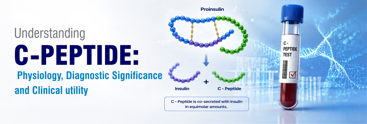
Understanding C – PEPTIDE: Physiology, Diagnostic Significance and Clinical utility

The C-peptide, also called the connecting peptide, is a 31-amino-acid polypeptide. It is secreted by pancreatic beta cells and is used to assess beta-cell function. It is cleaved from proinsulin to make insulin. It has a longer half-life than insulin and is therefore considered a useful and relatively stable indicator of pancreatic insulin secretion. It is also a marker of endogenous insulin secretion. C–peptide can help differentiate between endogenous insulin and exogenous insulin, as injected insulin cannot contain C-peptide.

Clinical Significance of C – PEPTIDE and Interpretation:

C-peptide levels are clinically important in the diagnosis and management of several conditions, including:

  • Assessment of pancreatic β-cell function:

C-peptide reflects the amount of insulin produced by the β-cell. It is particularly useful for determining residual β-cell function in patients. The key advantage is that C-peptide is a marker of endogenous insulin secretion. In contrast, an insulin measurement alone cannot consistently distinguish insulin produced by the pancreas from injected insulin.

  • Differentiating Diabetes Types

This test is used to differentiate between Type 1 and Type 2 Diabetes. Type 1 diabetes is identified by low C–peptide levels, and Type 2 diabetes is identified by high levels of C–peptide.

  • Chronic Kidney Disease (CKD)

As C–the kidneys excrete peptide, abnormal levels of it suggest Kidney dysfunction in the patient.

  • Hypoglycemia

It is a condition in which an individual’s blood glucose levels drop. It may be due to excessive insulin production. C–peptide test assists in identifying this condition.

  • It is very useful in the differential diagnosis of beta-cell tumors, insulinomas, and sulfonylurea therapy.

Different Diagnostic Ways to test for C-Peptide level:

Doctors recommend a C-Peptide test to measure the level of C-Peptide in human serum or plasma. Common laboratory methods for detecting C-Peptide levels include Chemiluminescence Immunoassay (CLIA), Enzyme-Linked Immunosorbent Assay (ELISA), Fluorescence Immunoassay (FIA), and Radioimmunoassay (RIA). The detection of C-Peptide levels is based on chemiluminescent immunoassay (CLIA) and is used for quantitative detection of C-Peptide in human serum and plasma.

Conclusion The C-peptide test is an important test for evaluating beta-cell function. C–peptide levels should always be interpreted alongside clinical history, glucose levels, HbA1c and other findings. Overall, C–peptide measurement provides important information for pancreatic function and can support more accurate diagnosis, classification and management of diabetes and other related disorders.

Explore the Product…

CA125: The Silent Signal—Detecting Ovarian Cancer before It Speaks

Cancer Antigen 125 (CA-125) is a glycoprotein biomarker used in the medical field for the detection and management of ovarian cancer. CA-125 is found in higher concentrations in the blood of women with ovarian cancer; its applications extend beyond cancer detection, as high levels may also be found in various non-cancerous conditions.

Clinical Significance of CA-125

CA-125 plays an important role in diagnosing and managing diseases, including:

  1. Ovarian Cancer Detection: Screening and Diagnosis with Monitored Treatment.
  2. Non-Ovarian Cancers: Elevated CA-125 levels are not exclusive to ovarian cancer. They are also observed in other malignancies, e.g., fallopian tube, endometrial, pancreatic, breast, and other colorectal cancers. In these cases, CA-125 is used more as a secondary marker for assessing the extent of disease or treatment effectiveness.
  3. Non-Cancerous Conditions:
  • Endometriosis: It is a condition in which tissue similar to the uterine lining grows outside the uterus, which raises the level of CA125.
  • Pelvic Inflammatory Disease (PID): This infection can also raise the level of CA125.
  • Menstruation
  • Pregnancy: During pregnancy, the CA-125 level rises in the first trimester.
  • Liver diseases like Cirrhosis raise the level of CA125.

Different Diagnostic ways to test for CA-125 level

The most common method for assessing CA-125 levels is a blood test.

Commonly used methods to detect CA-125 levels in laboratories include electrochemiluminescence (ECLIA), Chemiluminescence Immunoassay (CLIA), Enzyme-Linked Immunosorbent Assay (ELISA) and Radioimmunoassay (RIA).

The detection ofCA-125 based on the most advanced Chemiluminescence Immunoassay is used for quantitative measurement of Cancer Antigen 125 (CA125) in human serum/plasma as an aid in treatment monitoring of ovarian cancer.

Conclusion CA125 , a tumour marker, is primarily associated with epithelial ovarian cancer. While elevated levels of CA125 can indicate the presence of malignancy, particularly in postmenopausal women, higher levels are also observed in various benign conditions such as menstruation, endometriosis, pelvic inflammatory disease, and pregnancy. Therefore, in patients with confirmed ovarian cancer, CA125 is particularly useful for monitoring treatment response and detecting recurrence. Diagnosing CA125 levels through blood tests helps healthcare providers understand the underlying causes of ovarian cancer and develop appropriate treatment plans..

Explore the Product…

Advantages of Chemiluminescence in HIV, HCV, and HBsAg Testing

HIV, HCV, and HBsAg (HHH) detection in blood banks before transfusion is important for disease control, timely diagnosis, and prevention of their transmission, as these 3 markers are highly infectious. Chemiluminescence immunoassay CLIA have superior sensitivity, high specificity & rapid turnaround times compared to ELISA or radioimmunoassay. CLIA technology also facilitates accurate detection of analytes at very low concentrations, enhancing its performance in minimising false results and improving diagnostic accuracy. Additionally, its compatibility with automation and high-throughput testing makes chemiluminescence highly stable for regular clinical laboratories and blood screening.

Clinical Significance of Chemiluminescence in HHH range and Interpretation

  1. CLIA have excellent sensitivity for detecting both low and high concentrations of analytes in HIV, HCV & HBsAg, making it highly suitable for early diagnosis.
  2. Due to its low detection limits, CLIA can accurately identify very small amounts of analytes, which is important for detecting low-level HBsAg in clinical samples.
  3. Chemiluminescence technology plays a crucial role in infectious disease diagnosis in Blood banks to ensure safe blood transfusion and patient safety.
  4. CLIA tests for HIV and HBsAg screening are fourth-generation tests, as they detect both HIV antigens (p24) and antibodies and have better analytical sensitivity for HBsAg, thereby improving detection in early infection.

Interpretation of chemiluminescence results in HHH range:

The result interpretation is based on the sample-to-cutoff ratio (S/CO) in LIA. Since these are sensitive tests, borderline reactive samples should be carefully reanalysed through repeat testing and correlated with clinical symptoms and other diagnostic investigations for highly accurate & clinical interpretation.

Different diagnostic tests for HIV, HCV, and HBsAg (HHH)

Commonly used methods to detect HIV, HCV, and HBsAg in blood banks include electrochemiluminescence (ECLIA), Chemiluminescence Immunoassay (CLIA) & Enzyme-Linked Immunosorbent Assay (ELISA).

The 4th generation HIV chemiluminescence immunoassay, designed for the qualitative detection of antibodies to HIV-1 and HIV-2 and HIV-1 p24 antigen ,HBsAg CLIA for Hepatitis B surface antigen (HBsAg) and HCV CLIA for HCV Antibody detection is primarily used as a screening test for blood donors before transfusion to ensure blood safety.

Conclusion:

In conclusion, CLIA offers significant advantages over traditional ELISA and rapid tests for HIV, HCV & HBsAg screening, especially in terms of sensitivity, speed & automation in high-volume settings. CLIA utilises a luminescent molecule to emit light, providing an absolute measurement that results in superior analytical performance. CLIA also provides a wide linear range, allowing for precise detection of HHH markers compared to the limited absorbance range of ELISA. These methods also elevate workflow efficiency and decrease diagnostic errors, thereby ensuring safe blood transfusion in Blood banks and better patient management in Laboratories.

Explore the Product…

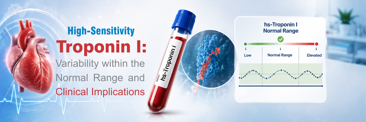
High-Sensitivity Troponin I: Variability Within the Normal Range and Clinical Implications

Troponin I (hs-TnI) is a key cardiac biomarker used for the prompt identification and diagnosis of myocardial injury. Troponin I, a protein involved in regulating cardiac muscle contraction, is released into the bloodstream when heart muscle cells are damaged.

Clinical Significance of High-Sensitivity Troponin I lies in its role in diagnosing and managing diseases, including:

  • Elevated hs-TnI is associated with a worse prognosis in chronic heart failure, chronic kidney disease, and diabetes.
  • May indicate ongoing subclinical myocardial injury.
  • May help rule out myocardial infarction in chest pain of non-cardiac origin.
  • Elevated hs-TnI may be observed in sepsis, pulmonary embolism, stroke, and COVID-19.

Different Diagnostic ways to test for hs–Troponin–I levels

Commonly used methods to detect hs-Troponin-I levels in laboratories include electrochemiluminescence (ECLIA), Chemiluminescence Immunoassay (CLIA), Enzyme-Linked Immunosorbent Assay (ELISA), and fluorescence-based immunoassay methods (FIA). The detection of hs-Troponin-I, based on a Chemiluminescence Immunoassay, is used for quantitative measurement of hs-Troponin-I levels in human serum/plasma as an aid in the diagnosis of Myocardial Infarction.

Conclusion Highly Sensitive Troponin I is a valuable cardiac biomarker for accurately detecting myocardial injury, including minor myocardial damage.  Increased hs-TnI levels are commonly detected in more severe cases and are closely linked to higher rates of complications, including myocarditis, arrhythmias, heart failure, and overall mortality. Its high sensitivity is useful for early risk assessment and treatment planning.

Explore the Product…

D-Dimer:  Biomarker for Diagnosis of Thrombotic Disorders

Introduction

D-dimer is a protein fragment formed when cross-linked fibrin is broken down by plasmin during fibrinolysis. It is present in very low concentrations in the bloodstream. Under normal physiological conditions, clot formation and destruction remain balanced, preventing excess bleeding and ensuring proper blood flow after blood vessel healing.

Elevated D-dimer levels are found in disseminated intravascular coagulation (DIC), deep vein thrombosis (DVT), and pulmonary embolism (PE). Other circumstances, such as old age, pregnancy, cancer, liver disease, and infection, may also lead to high D-dimer levels. The ELISA system has higher sensitivity than latex agglutination tests, providing improved aid in the diagnosis and exclusion of venous thromboembolism (VTE).

Clinical Significance of D-Dimer

D-dimer plays an essential role in diagnosing, managing, and monitoring several medical conditions involving thrombosis and coagulation disorders.

1. Diagnosis of Venous Thromboembolism (VTE)

D-dimer testing is extensively used to rule out venous thromboembolism, including:

  • Deep Vein Thrombosis (DVT)
  • Pulmonary Embolism (PE)

2. Disseminated Intravascular Coagulation (DIC)

D-dimer concentrations increase significantly in patients with DIC due to widespread activation of coagulation and fibrinolysis. It is an important laboratory marker included in DIC diagnostic scoring systems.

3. Cardiovascular Diseases

Elevated D-dimer levels have been found in Patients diagnosed with cardiovascular disorders, including acute coronary syndrome, ischemic stroke, peripheral arterial disease, and atrial fibrillation.

4. COVID-19 and Critical illness

During COVID-19 pandemic, D-dimer was recognised as a valuable prognostic marker . Elevated D-dimer levels were frequently associated with severe disease, thromboembolic complications, the need for intensive care, and an increased risk of mortality. Consequently, D-dimer testing became an integral part of clinical assessment, helping clinicians monitor disease progression and identify patients at greater risk of adverse outcomes.

5. Other Clinical Applications

Elevated D-dimer levels are also observed in malignancies, pregnancy, liver disease, trauma, major surgery, sepsis, and advanced age.

Different Methods for D-Dimer Diagnosis

Doctors recommend D-dimer testing to measure D-dimer levels in serum or plasma. Normal D-dimer levels vary depending on factors such as the individual’s age and sex. For a healthy human body, the reference range is generally less than 500 ng/mL (nanograms per millilitre) or less than 0.50 mg/L (milligrams per litre).

Conclusion

D-dimer is a vital biomarker for evaluating thrombotic and fibrinolytic activity in patients with suspected coagulation disorders. Elevated D-dimer levels are not specific for any single disease; they provide critical diagnostic and prognostic information when interpreted in conjunction with clinical assessment and imaging studies. Various laboratory methods—including ELISA, latex agglutination, immunoturbidimetric assays, fluorescence immunoassays, and CLIA—are available for D-dimer estimation, each with different advantages and limitations. The chemiluminescence-based tests are a better diagnostic approach due to higher accuracy and excellent reproducibility. The advancement of CLIA-based diagnostic platforms is expected to enhance the clinical utility of D-dimer testing further and improve patient outcomes across a broad range of medical specialities.

Explore the Product…

Beyond Diet: The Truth About Celiac Disease

Introduction

Celiac disease occurs in individuals as a reaction to gluten sensitivity. Gluten is a protein that is found in various cereals like Wheat, Barley, and rye. If a patient with celiac disease consumes food containing gluten, this will lead to damage to the mucous membrane of the small intestine.

Tissue transglutaminase (tTG) is an enzyme that is released from cells during inflammation. Measurement of serum anti-tissue transglutaminase (tTG) antibodies is the most common serological test used in screening for celiac disease. To treat celiac disease, doctors advise patients to eat gluten-free food.

Clinical Significance of anti-tTG and Interpretation

Celiac disease has significant clinical importance because it can affect multiple organ systems. If left undiagnosed or untreated, celiac disease may lead to serious complications such as malnutrition, anaemia, osteoporosis, neurological disorders, growth retardation and an increased risk of some intestinal malignancies. Early identification of disease and associated risk factors plays a crucial role in timely diagnosis, disease prevention, and long-term patient management.

The interpretation of genetic and risk-factor analysis in celiac disease requires careful clinical correlation with serological tests, symptoms, and the person’s diet.

Different Diagnostic ways to test for anti-tTG IgA antibody levels

A doctor recommends an anti-tTG IgA antibody test to check for Celiac Disease.

Commonly used methods to detect Anti-tTG IgA antibody levels in laboratories include electrochemiluminescence (ECLIA), immunoturbidimetric method, Chemiluminescence Immunoassay (CLIA), Enzyme-Linked Immunosorbent Assay (ELISA) and Fluorescent Enzyme Immunoassay (FEIA).

The detection of anti-tTG IgA Antibodies is based on the most advanced Chemiluminescence Immunoassay. It is used for qualitative detection of anti-tTG IgA antibodies in Human Serum or Plasma samples as an aid in recognition and diagnosis of Celiac disease.

Conclusion

Celiac Disease is an autoimmune disorder that is affected by factors such as family history, gluten exposure, infections, and other autoimmune conditions, which contribute to Celiac disease development and progression. Ultimately, Early identification of the disease plays a crucial role in timely diagnosis, disease prevention, and long-term patient management. The interpretation of genetic and risk-factor analysis in celiac disease requires careful clinical correlation with serological tests, symptoms, and the person’s diet.

Explore the Product…

Progesterone: The hormone that ensures balance, harmony, and a healthy pregnancy

Progesterone is an essential hormone that regulates multiple biological processes in the female reproductive system. It is produced primarily in the ovaries, adrenal glands, and placenta during pregnancy. Progesterone has an essential function in preparing the endometrium layer of the uterus for Implantation. In addition to its fertility functions, progesterone influences other bodily systems, such as the brain and cardiovascular health.

Clinical Significance of Progesterone and Interpretation:

Progesterone plays a vital role in conditions including:

  • Maintenance of the uterus during pregnancy.
  • Treatment of secondary amenorrhea and endometrial hyperplasia.
  • Marker for fertility; low levels lead to lowered fertility.
  • To monitor conditions like ectopic pregnancy (a condition in which a fertilised egg implants other than the uterus, such as the fallopian tube), miscarriage, and cancers like adrenal and Ovarian.
  • To monitor PCOS – polycystic ovarian syndrome, as progesterone levels become higher in the presence of Cysts.
  • To determine the cause of abnormal bleeding in the uterus.

Diagnostic methods available for testing progesterone levels:

A doctor recommends a progesterone test to measure progesterone levels in the blood. Commonly used methods for detecting progesterone levels in laboratories include: Chemiluminescence Immunoassay (CLIA), chromatographic techniques such as High-Performance Liquid Chromatography (HPLC), Radioimmunoassay (RIA), Enzyme-Linked Immunosorbent Assay (ELISA), and Progesterone Receptor Assay.

The detection of Progesterone based on the most advanced chemiluminescence immunoassay is used for quantitative measurement of Progesterone level in human serum/plasma samples as an aid in the diagnosis of threatened abortion in conjunction with other laboratory and clinical findings.

Conclusion Progesterone  plays a vital role in regulating many body functions, particularly in women. It helps regulate the menstrual cycle, supports a healthy pregnancy, and contributes to overall hormonal balance. When progesterone levels become too low or too high, they can disrupt normal body functions and lead to a range of health concerns.

Explore the Product…

CK-MB: An Important Cardiac Biomarker for the Diagnosis of Heart Muscle Damage

CK-MB is a cardiac marker for the diagnosis of myocardial infarction and myocarditis. It is a creatine kinase enzyme composed of muscle and brain subunits and can be used to assess tissue injury. When the myocardium experiences structural injury or oxygen deprivation, it releases CK-MB directly into the bloodstream. Previously, CK-MB was considered the standard for diagnosing a heart attack; however, now HS Troponin I is considered the gold standard. CK-MB plays a crucial role in some cases, such as detecting Reinfarction. It helps diagnose even minor irritations as well as severe complications. In people with advanced kidney disease, doctors sometimes use CK-MB to get a clear timeline of acute events.

Clinical Significance of CK-MB and Interpretation:

Abnormal CK-MB levels are clinically important in diagnosing and managing several conditions, including:

  • Acute Myocardial infarction: CK-MB levels rise approximately 3-6 hours after myocardial infarction, peak in 12-24 hours and return to normal within 48-72 hours.
  • Reinfarction: . When chest pain accompanies elevated CK levels/ CK-MB along with chest pain ,it often indicates a recent heart attack. If CK-MB levels initially rise and then fall, followed by another increase, this may suggest a second heart attack or ongoing heart damage.
  • Monitoring Post–Heart Procedures: It helps detect even minor irritations and severe complications after angioplasty, stent placement, and CABG (Coronary artery bypass graft) surgery.

Different Diagnostic Ways to test for CK-MB level:

Doctors recommend a CK-MB test to measure the level of CK-MB in human serum or plasma.

Common laboratory methods for detecting CK-MB levels include Chemiluminescence Immunoassay (CLIA), Enzyme-Linked Immunosorbent Assay (ELISA), and Fluorescence Immunoassay (FIA). CK-MB detection is based on the most commonly used chemiluminescent immunoassay (CLIA) for quantitative measurement in human serum and plasma.

Conclusion

Measuring CK-MB is crucial for diagnosing myocardial infarction, particularly in the case of Acute Myocardial Infarction (AMI) as it is present in heart muscle cells. CK-MB levels typically rise 4-6 hours after a myocardial infarction, peak at around 24 hours, and return to normal within three days. This pattern of elevation and decline, combined with other cardiac markers, ECG changes, and the patient’s chest pain history, has become an essential method for diagnosing acute myocardial infarction.

Explore the Product…

Prostate-Specific Antigen (PSA) – Early Detection, Lifesaving Insights

Prostate-Specific Antigen (PSA) is produced by the prostate gland, located in the male reproductive system. There are two forms of PSA  ; total PSA ( bound to proteins ) and free PSA (freely circulating) . The ratio of free PSA to total PSA ( per cent-free PSA) is usually found to be comparatively low in patients  with prostate cancer compared to those without the condition.

PSA as a Diagnostic Biomarker

The prostate-specific antigen (PSA) test is widely used to evaluate prostate health and plays an important role in detecting prostate cancer. PSA test results provide valuable information in combination with other diagnostic methods, such as imaging scans or a prostate biopsy. PSA testing is also used to monitor whether the cancer has recurred or if the patient is in remission.

Factors That Influence PSA Levels

Age: PSA levels increase slightly with age.

Benign prostatic hyperplasia (BPH): PSA levels may increase in this condition (non-cancerous enlargement of the prostate).

Medical Procedures: Medical procedures, such as prostate biopsies or catheterisation, can raise PSA levels temporarily.

Different Diagnostic Ways to test for PSA level

  • Measurement of PSA levels in the blood sample to screen for prostate cancer.
  • Serum PSA Level – Most men have PSA levels under 4 ng/mL, which means they are not affected by prostate cancer.
  • Commonly used methods to detect PSA Levels in laboratories include Chemiluminescence Immunoassay (CLIA), Enzyme-Linked Immunosorbent Assay (ELISA), Radioimmunoassay (RIA), Magnetic Resonance Imaging, Prostate-Specific Risk Stratification Bioassays, and fluorescent immunoassay.
  • The detection of PSA using the most advanced Chemiluminescence Immunoassay is used for quantitative measurement of Prostate-specific antigen (t-PSA) in human serum/plasma, and for monitoring in clinical observation, prognosis, and recurrence monitoring of prostate cancer.

Conclusion:  PSA levels are important for both patients and healthcare providers to make informed decisions. Monitoring of PSA tests, especially for men at higher risk for prostate cancer, can ensure that individuals take a proactive approach to their prostate health.

Explore the Product…